anatomical human muscular skeleton model
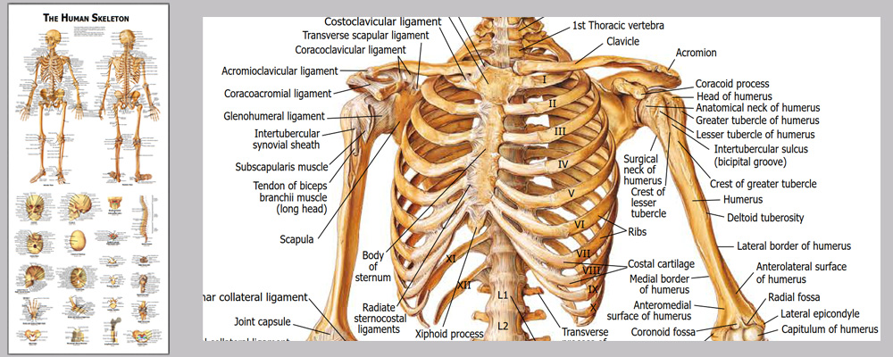
This life size articulated adult human skeleton model is 180cm tall & ideal for teaching / learning the basics of human anatomy. Includes a colorful Human Skeleton chart to show all the detailed structures for reference.Detailed colorful chart with over 630 accurate definitions. Durable and no reflection with matte film covered, 36 * 78cm

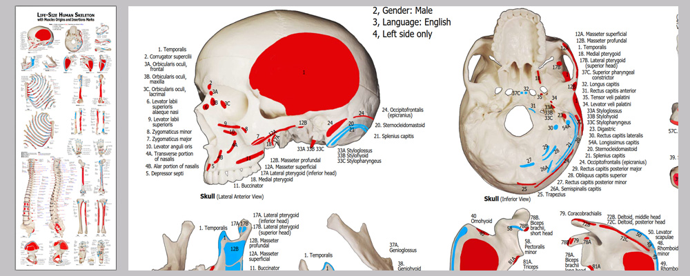
Detailed muscles origins and insertions with codes. Durable and no reflection with matte film covered, 100cm * 38cm

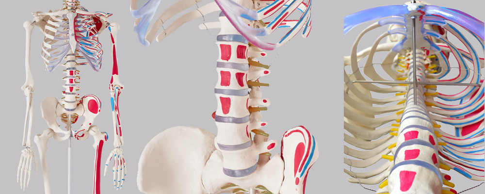
Stainless steel wires keep the ribs gaps stable.

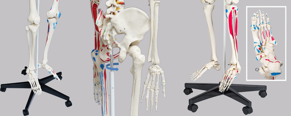
2 of 5 casters are lockable.

An overview of JC anatomy advantages: • Hand-painted muscle origins and insertions • Flexible spine and ligaments • Slipped disc between the 3rd and 4th lumbar vertebrae • Protruding spinal nerves and vertebral arteries • 3-part assembled skull with individually inserted teeth • Made from a durable, unbreakable synthetic material • Top quality, life size natural casting • lMultiple Applications -photography,chiropractor,chiropractor, etc. • lChart - Detailed colorful chart with about 711 accurate definitions. Durable and no reflection with matte film covered, 36 * 78cm • On a stable metal stand with 5 casters (painted white) • Full size dust cover keeps model clean while in storage • Exceptional value for money with a 3 year guarantee • anatomical human skeleton model Easy to use - Main joints are movable. Skull, skullcap arms, legs, crus, feet are removable.
| Product name | anatomical human skeleton model |
| Place of Origin | Shenzhen China |
| Product Material | PVC, ABS, SST |
| Rib cage | A 5mm dia |
| Human skeleton model life | 19years |
| Surface treatment | Polish. Etched. Texture |
| SUPPORT 24/7 | Contact us 24 hours a day, 7 days a week |
| Size | 77 * 43 * 137 |
| Port | Shenzhen |
| PAYMENT & ORDERING | PayPal account or pay by credit card |
anatomical human skeleton model FAQs Guide Are you looking for a quick review guide about anatomical human skeleton model? An ultimate FAQs buying guide is available to help you.This guide contains all the information about all the important facts, figures, and various processes regarding anatomical human skeleton model. Let’s continue!
2.About anatomical human skeleton model warranty
3.anatomical human skeleton model Are accompanying labels provided indicating the name and location of each bone?
4.anatomical human skeleton model Can it be used to show the differences in body structure between children and adults?
5.anatomical human skeleton model Can it be used as a teaching aid?
6.Does the anatomical human skeleton model have movable joints that can show the range of motion of various parts of the human body?
7.How similar is the anatomical human skeleton model appearance to a real human skeleton?
8.How many movable joints does anatomical human skeleton model have?
9.Can the anatomical human skeleton model be disassembled and assembled to facilitate learning and display?
10.How can a anatomical human skeleton model be utilized for rehabilitation and physiotherapy purposes?
11.Can the anatomical human skeleton model display many different body poses?
12.Is it possible to disassemble different parts of this anatomical human skeleton model?
13.Are anatomical human skeleton model joints elastic and flexible?
14.Can anatomical human skeleton model be used in medical research?
15.How to correctly assemble and utilize a anatomical human skeleton model?
1.How durable is anatomical human skeleton model?
The durability of a human skeleton model can vary depending on the materials used and the quality of construction. However, generally speaking, a high-quality skeleton model can be quite durable and can last for many years if properly cared for. Here are some factors that can affect the durability of a human skeleton model: 1. Materials: The most common materials used for human skeleton models are plastic, resin, and wood. Plastic and resin models are generally more durable as they are less prone to damage from impacts or moisture. Wood models can also be quite durable, but they may be more susceptible to wear and tear over time. 2. Maintenance: Proper maintenance and care can significantly increase the durability of a human skeleton model. Regular cleaning with a soft cloth and mild detergent can help keep the model free of dust and dirt. Additionally, storing the model in a protective case or covering can also help prevent damage. Overall, a high-quality human skeleton model can be quite durable and can last for many years with proper care. However, it is important to keep in mind that no model is completely indestructible, and regular wear and tear can occur over time.
2.About anatomical human skeleton model warranty
3 years. If any problem occurs with the model due to material or structural defects, it will be repaired or replaced free of charge to the buyer
3.anatomical human skeleton model Are accompanying labels provided indicating the name and location of each bone?
The stardard model does not provide this function. The chart shipped with pro model shows the name and location of each bone
4.anatomical human skeleton model Can it be used to show the differences in body structure between children and adults?
Yes, a human skeleton model can be used to clearly demonstrate the differences in body structure between children and adults. Here are some specific examples of how a skeleton model can be used to illustrate these differences: Size and proportion: One of the most evident differences between children and adults is their size and proportion. A skeleton model can be used to compare the relative sizes of different bones in a child and adult, such as the skull, spine, and limbs. Children have smaller and more delicate bones compared to adults, and their proportions also differ. For example, the head and skull of a child are proportionally larger to their body compared to an adult. In conclusion, a human skeleton model is an effective educational tool to showcase the differences in body structure between children and adults. Its three-dimensional design and ability to highlight specific bone structures make it an excellent visual aid to help understand the changes that occur during growth and development.
5.anatomical human skeleton model Can it be used as a teaching aid?
Yes, a human skeleton model is a commonly used teaching aid in various academic fields such as anatomy, biology, and medical education. It is an accurate and detailed representation of the human skeleton, providing a visual aid for students to understand and grasp the complexities of the skeletal system.
6.Does the anatomical human skeleton model have movable joints that can show the range of motion of various parts of the human body?
Yes, the human skeleton model typically has movable joints that can show the range of motion of various parts of the human body. These joints are usually represented by different types of articulations such as ball and socket, hinge, saddle, pivot, and gliding joints, which mimic the movement of the actual joints in the human body. For example, the shoulder joint of a human skeleton model will have a ball and socket structure that allows for a wide range of motion, similar to the shoulder joint in a real human body. This joint is able to rotate 360 degrees and can also move forward, backward, and sideways. Similarly, the elbow joint in a human skeleton model will have a hinge structure that allows for flexion and extension movements, just like the elbow joint in a real human body. This enables the model to accurately demonstrate movements such as bending and straightening of the arm.
7.How similar is the anatomical human skeleton model appearance to a real human skeleton?
depending on the level of detail and accuracy that is represented. In general, a well-made human skeleton model will closely resemble a real human skeleton in terms of size, shape, and overall structure. The bones of a human skeleton model are typically made of a strong and durable material such as plastic or resin, and are molded and assembled to accurately represent the bone structure of a real human body. The proportions and dimensions of the bones are also generally accurate, with consideration given to the size and shape of bones in different areas of the body. In addition to the basic bone structure, a human skeleton model may also feature more detailed elements such as muscle attachments and articulations. These details help to further enhance the realism of the model and make it more closely resemble a real human skeleton.
8.How many movable joints does anatomical human skeleton model have?
There are approximately 13 movable joints in the human skeleton model, with different types of joints including ball and socket, hinge, pivot, gliding, and saddle joints. These joints allow for a wide range of movements, such as flexion, extension, rotation, abduction, and adduction.
9.Can the anatomical human skeleton model be disassembled and assembled to facilitate learning and display?
Yes, many human skeleton models are designed to be disassembled and assembled for educational purposes. The bones are typically connected by metal or plastic clips that can be easily removed and reattached, allowing for the skeleton to be taken apart and put back together. This feature makes the model useful for learning about the different bones and their connections, as well as their placement in the body. Students can learn the names and locations of each bone by taking them apart and reassembling them in the correct order. This hands-on approach can be helpful for visual learners and can make the learning process more engaging and interactive.
10.How can a anatomical human skeleton model be utilized for rehabilitation and physiotherapy purposes?
Demonstrate the right bone parts postion relationships can help doctors and patients to understanhuman skeleton model can be a valuable tool for rehabilitation and physiotherapy purposes as it can aid in teaching and demonstrating proper body mechanics, identifying and addressing musculoskeletal imbalances and dysfunctions, and monitoring progress in treatment. Here are some ways in which a human skeleton model can be utilized for rehabilitation and physiotherapy purposes: 1. Education: A human skeleton model can be used to educate patients and clients about the structure and function of the human body. This can help them understand their injuries or conditions better, and also make them more aware of how their body moves and functions. 2. Creating personalized treatment plans: With the help of a skeleton model, therapists can design personalized treatment plans for each individual patient. This can include exercises and movements targeted at specific areas of the body that need strengthening or stretching.d the right way of rehabilitation and physiotherapy.
11.Can the anatomical human skeleton model display many different body poses?
The Human skeleton model can display many different body poses in order to show the movement and positioning of bones and muscles. The model can be manipulated into a range of poses such as the classical anatomical position, where the arms are down by the sides, palms facing forward, and feet shoulder-width apart. It can also be posed to demonstrate actions such as walking, running, jumping, and sitting. The flexibility of the model allows for a wide range of movements to be shown, from simple movements of individual joints to complex combinations of actions involving multiple joints. Additionally, the model can display different body poses by adjusting the positions of individual bones and joints. For example, the arms can be raised or lowered, the elbows bent or straightened, and the fingers spread or closed. The legs can be straightened or bent at the knee, and the ankles can be rotated to show different directions of movement. These adjustments allow for a variety of poses to be demonstrated, from relaxed positions to more dynamic and active positions. In summary, the Human skeleton model can display a wide variety of body poses and movements, ranging from simple to complex, to showcase the functions and interactions of the bones and muscles in the human body.
12.Is it possible to disassemble different parts of this anatomical human skeleton model?
It's easy to disassemble the whole model into some sub-assemblies - Skull, torso, extremities, etc toollessly. The model can be disassembled into more pieces with tools (Not recommended)
13.Are anatomical human skeleton model joints elastic and flexible?
The human skeleton model joints are not entirely elastic and flexible, as they include various types of joints with different levels of flexibility. The elasticity and flexibility of a joint depend on its structure and function.
14.Can anatomical human skeleton model be used in medical research?
This model is mainly for displaying the skeleton structure, shapes, position relationship, and simulating joints movements, surface texture of the bones.
15.How to correctly assemble and utilize a anatomical human skeleton model?
When assembling and utilizing a human skeleton model, there are several steps to follow to ensure correct positioning and accurate representation of the skeletal system. Below is a step-by-step guide to properly assemble and utilize a human skeleton model: 1. Gather all the necessary components: The first step is to gather all the pieces of the human skeleton model, which typically includes a skull, rib cage, spinal column, arms, and legs. Make sure that all the pieces are present and in good condition. 2. Identify the bones: Before assembling the skeleton, it’s important to familiarize yourself with the different bones and their names. The skull, for example, has different parts such as the cranium, mandible, and maxilla, while the spinal column consists of the cervical, thoracic, lumbar, sacrum, and coccyx vertebrae. 3. Attach the arms and legs: Next, attach the arms and legs to the skeleton. Start by connecting the arms to the shoulder sockets and then attach the hand bones to the arms using the elbow and wrist joints. For the legs, connect the femur (thigh bone) to the hip socket, followed by the tibia and fibula (lower leg bones), and finally the foot bones.


