human anatomy skeleton model
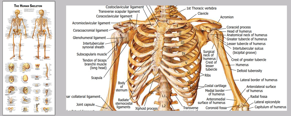
This life size articulated adult human skeleton model is 180cm tall & ideal for teaching / learning the basics of human anatomy. Includes a colorful Human Skeleton chart to show all the detailed structures for reference.Detailed colorful chart with over 630 accurate definitions. Durable and no reflection with matte film covered, 36 * 78cm

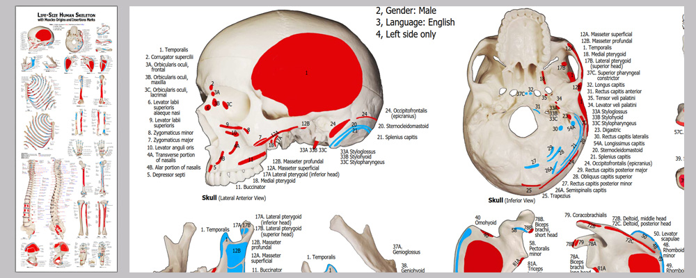
Detailed muscles origins and insertions with codes. Durable and no reflection with matte film covered, 100cm * 38cm

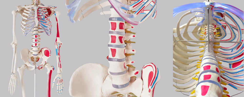
Stainless steel wires keep the ribs gaps stable.

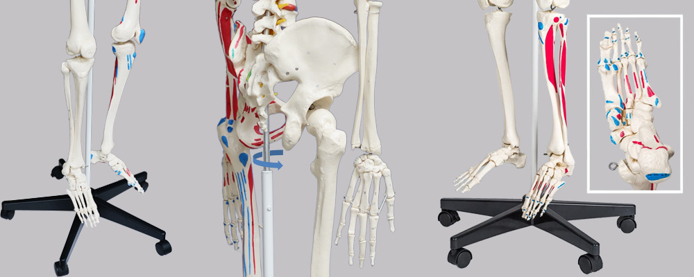
2 of 5 casters are lockable.

An overview of JC anatomy advantages: • Hand-painted muscle origins and insertions • Flexible spine and ligaments • Slipped disc between the 3rd and 4th lumbar vertebrae • Protruding spinal nerves and vertebral arteries • 3-part assembled skull with individually inserted teeth • Made from a durable, unbreakable synthetic material • Top quality, life size natural casting • lMultiple Applications -Physiology,Physiology,Physiology, etc. • lChart - Detailed colorful chart with about 733 accurate definitions. Durable and no reflection with matte film covered, 36 * 78cm • On a stable metal stand with 5 casters (painted white) • Full size dust cover keeps model clean while in storage • Exceptional value for money with a 3 year guarantee • human anatomy skeleton model Easy to use - Main joints are movable. Skull, skullcap arms, legs, crus, feet are removable.
| Product name | human anatomy skeleton model |
| Place of Origin | Shenzhen China |
| Product Material | PVC, ABS, SST |
| Rib cage | A 5mm dia |
| Human skeleton model life | 15years |
| Surface treatment | Polish. Etched. Texture |
| SUPPORT 24/7 | Contact us 24 hours a day, 7 days a week |
| Size | 82 * 24 * 137 |
| Port | Shenzhen |
| PAYMENT & ORDERING | PayPal account or pay by credit card |
human anatomy skeleton model FAQs Guide Are you looking for a quick review guide about human anatomy skeleton model? An ultimate FAQs buying guide is available to help you.This guide contains all the information about all the important facts, figures, and various processes regarding human anatomy skeleton model. Let’s continue!
2.Can bone diseases such as osteoporosis be displayed on the human anatomy skeleton model?
3.Is it possible to disassemble different parts of this human anatomy skeleton model?
4.Does this human anatomy skeleton model include all human skeletal systems?
5.Does the human anatomy skeleton model have movable joints that can show the range of motion of various parts of the human body?
6.Can human anatomy skeleton model be used in medical research?
7.About human anatomy skeleton model production capacity
8.How many thoracic and lumbar vertebrae are there in the human anatomy skeleton model?
9.Can the human anatomy skeleton model be disassembled and assembled to facilitate learning and display?
1.How durable is human anatomy skeleton model?
The durability of a human skeleton model can vary depending on the materials used and the quality of construction. However, generally speaking, a high-quality skeleton model can be quite durable and can last for many years if properly cared for. Here are some factors that can affect the durability of a human skeleton model: 1. Materials: The most common materials used for human skeleton models are plastic, resin, and wood. Plastic and resin models are generally more durable as they are less prone to damage from impacts or moisture. Wood models can also be quite durable, but they may be more susceptible to wear and tear over time. 2. Maintenance: Proper maintenance and care can significantly increase the durability of a human skeleton model. Regular cleaning with a soft cloth and mild detergent can help keep the model free of dust and dirt. Additionally, storing the model in a protective case or covering can also help prevent damage. Overall, a high-quality human skeleton model can be quite durable and can last for many years with proper care. However, it is important to keep in mind that no model is completely indestructible, and regular wear and tear can occur over time.
2.Can bone diseases such as osteoporosis be displayed on the human anatomy skeleton model?
Yes, bone diseases like osteoporosis can be displayed on a human skeleton model. A human skeleton model is a representation of the bones and joints of the human body and can be used to educate and train students and medical professionals. Osteoporosis is a bone disease that results in weakened and porous bones, increasing the risk of fractures. As a result, there are certain features and changes in the bone structure that can be seen on a human skeleton model.
3.Is it possible to disassemble different parts of this human anatomy skeleton model?
It's easy to disassemble the whole model into some sub-assemblies - Skull, torso, extremities, etc toollessly. The model can be disassembled into more pieces with tools (Not recommended)
4.Does this human anatomy skeleton model include all human skeletal systems?
It depends on the model in question. Some Human skeleton models may include all human skeletal systems such as the axial skeleton (skull, vertebral column, ribcage), appendicular skeleton (arms, legs, shoulder girdle, pelvic girdle), and cartilaginous skeleton (cartilaginous structures such as the nose and ears). However, others may only include certain systems or be limited to specific areas of the body. It is important to carefully read the product description or ask the manufacturer for a detailed list of what is included in the model to ensure that it meets your needs.
5.Does the human anatomy skeleton model have movable joints that can show the range of motion of various parts of the human body?
Yes, the human skeleton model typically has movable joints that can show the range of motion of various parts of the human body. These joints are usually represented by different types of articulations such as ball and socket, hinge, saddle, pivot, and gliding joints, which mimic the movement of the actual joints in the human body. For example, the shoulder joint of a human skeleton model will have a ball and socket structure that allows for a wide range of motion, similar to the shoulder joint in a real human body. This joint is able to rotate 360 degrees and can also move forward, backward, and sideways. Similarly, the elbow joint in a human skeleton model will have a hinge structure that allows for flexion and extension movements, just like the elbow joint in a real human body. This enables the model to accurately demonstrate movements such as bending and straightening of the arm.
6.Can human anatomy skeleton model be used in medical research?
This model is mainly for displaying the skeleton structure, shapes, position relationship, and simulating joints movements, surface texture of the bones.
7.About human anatomy skeleton model production capacity
600pcs each month
8.How many thoracic and lumbar vertebrae are there in the human anatomy skeleton model?
There are 12 segments in the thoracic vertebrae, 5 segments in the lumbar vertebraeGenerally, there are 12 thoracic vertebrae and 5 lumbar vertebrae in the human skeleton model. The thoracic vertebrae are located in the middle region of the spine, between the cervical vertebrae (neck) and lumbar vertebrae (lower back). They are numbered from T1 to T12, with T1 being the first thoracic vertebra closest to the skull and T12 being the last thoracic vertebra closest to the pelvis. The lumbar vertebrae are located in the lower back region, below the thoracic vertebrae. They are numbered from L1 to L5, with L1 being the first lumbar vertebra closest to the thoracic vertebrae and L5 being the last lumbar vertebra closest to the sacrum (a triangular bone at the base of the spine). However, the specific number of thoracic and lumbar vertebrae may vary slightly from person to person due to individual differences in bone structure. Some people may have 11 or 13 thoracic vertebrae, and 4 or 6 lumbar vertebrae. This is known as a variation or anomaly in the number of vertebrae. .
9.Can the human anatomy skeleton model be disassembled and assembled to facilitate learning and display?
Yes, many human skeleton models are designed to be disassembled and assembled for educational purposes. The bones are typically connected by metal or plastic clips that can be easily removed and reattached, allowing for the skeleton to be taken apart and put back together. This feature makes the model useful for learning about the different bones and their connections, as well as their placement in the body. Students can learn the names and locations of each bone by taking them apart and reassembling them in the correct order. This hands-on approach can be helpful for visual learners and can make the learning process more engaging and interactive.


